Encyclopedia entry
Ulnar Artery
Abstract
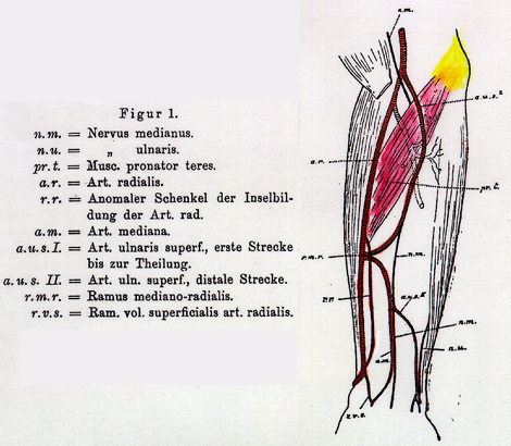
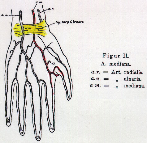
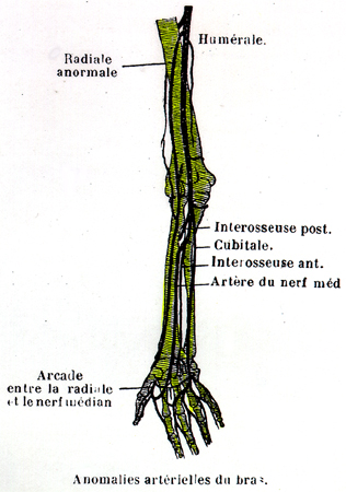
The ulnar artery may arise from the brachial above the usual point of division or from the axillary, in which case it usually passes over the flexor muscles, but beneath the fascia, to reach its usual position in the forearm. In circumstances such as this, the artery may terminate in the proximal forearm with a persistent median artery supplying the superficial arch The recurrent arteries and the common interosseous are usually derived from the trunk vessel from which the ulnar arises.
The ulnar artery may arise from the brachial above the usual point of division or from the axillary, in which case it usually passes over the flexor muscles, but beneath the fascia, to reach its usual position in the forearm. In circumstances such as this, the artery may terminate in the proximal forearm with a persistent median artery supplying the superficial arch
The recurrent arteries and the common interosseous are usually derived from the trunk vessel from which the ulnar arises.
It may run beneath the muscles or merely beneath the skin, where it becomes vulnerable to iatrogenic or self-inflicted problems.
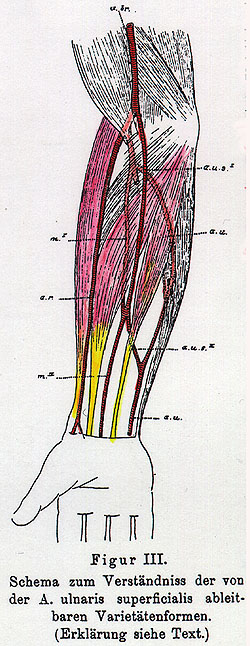
In cases of high division of the brachial, the ulnar may run in a superficial, subcutaneous position or beneath the fascia throughout its extent in the forearm.
In some cases of high origin and superficial course, it gives rise to the subscapular and profunda brachii but not the common interosseous.
It may also provide an accessory ulnar recurrent, radial recurrent, or accessory interosseous branch, which may enter the palm and provide any of the branches of the superficial palmar arch.
In some cases in which the origin of the brachial is normal, the ulnar takes a superficial course, covered by fascia in the forearm, and the recurrent branches and the common interosseous arise from the radial.
The ulnar may be a branch of profunda brachii.
The ulnar is sometimes doubled; in some cases one of the two arteries may take a superficial course in the forearm.
At the wrist, the ulnar artery lies on the flexor retinaculum accompanied by the ulnar nerve at the ulnar side of the artery. It then lies to the radial side of the pisiform bone and to the ulnar side of the hook of the hamate, the two bones form a protective channel (of Guyon) for artery and nerve that is converted into a canal by the flexor retinaculum. The artery and nerve may be compromised if a variation of the abductor digiti minimi also occupies the canal.
The ulnar artery is more variable in its position than the radial artery.
R. Quain describes the variations in the Ulnar artery as follows: In the whole of the cases observed, the ulnar artery was found to deviate from its usual mode of origin nearly in proportion of one in 13 (~7.7%). The brachial artery was, more frequently than the axillary the source from which it sprang; and the lower part of the brachial than the upper. In one case of high origin of the ulnar artery, the vessel was connected with the brachial opposite the elbow-joint by means of a transverse branch (read also remarks on the variations of the axillary and brachial arteries). The course and position of the ulnar artery in the forearm is more frequently variable than that of the radial.
In cases of high origin it almost invariably descends over the muscles arising from the inner (medial) condyle of the humerus, only one exception was found by R. Quain. In one instance the ulnar artery taking this course divided just below the elbow into a superficial and deep branch.
Most commonly in such cases it is covered by the fascia of the forearm; but instances also occur in which the artery rests on the fascia, and either continues in that position or again sinks below the fascia distally, while the vessel thus disposed is distributed distally after the manner of the usual ulnar artery. The vessel from which the high ulnar separates is afterwards divided into the radial and the interosseous; it appears therefore probable the this unusual arrangement results from the early obstruction of the ulnar artery below the origin of the interosseous artery, and the development of a superficial vas aberrans, which unites that portion of the vessel below the obstruction with the axillary or brachial trunk. The interosseous artery in such cases of variability thus comprises not only the ordinary interosseous branch, but also the portion of the ulnar artery above the obstruction; and, in accordance with this view (Quain's), one finds that the recurrent branches are derived from it.
The ulnar artery has been found taking a superficial course when arising in the usual situation, and in these cases also the reccurent and interosseous arteries may be given off by the radial trunk.
Heuston reported an ulnar artery, arising from the brachial at its usual place of origin, passed superficial to the flexor muscles of the forearm, giving off no important branch. The radial artery, while in the antecubital fossa, gave off a large branch, from which the following vessels had their origin- viz., anterior and posterior ulnar recurrent, radial recurrent, comes nervi mediani, and anterior and posterior interosseous, all of which had a normal distribution. High Origin (HO) of the Arteries of the Forearm Ulnar Artery
Number of Observations
HO Total
Origin 1) 380 112
Axillary -
Brachial
Upper third -
Middle third -
Lower third - 2) 270 60
Axillary
21
Brachial
Upper third 15
Middle third 16
Lower third 8 3) 136 26
Axillary 7
Brahcial
Upper third 7
Middle third 6
Lower third 9
HO= High Origin
1) Statistic sby Giacomini. 2) Statistics by authors cited by Giacomini. 3) Statistics by Dubreuil-Chambardel.
Precocious or high origin of the ulnar artery is reported by Dubreuil-Chambardel to be very frequent, ~24%. High Origin (HO) of the Arteries of the Forearm Radio-Ulnar Trunk
Number of Observations
HO Total
Origin 1) 380 22 2) 270 25
Axillary 12
Brachial 13 3) 136 5
Axillary 2
Brachial 3
1) Statistics by Giacomini. 2) Statistics by authors cited by Giacomini. 3) Statistics by Dubreuil-Chambardel. High Origin of the Arteries of the Forearm Radio-Ulnar-Interosseous Trunk
Number of Observations Total Origin:
1.
0
2.
270 24 ?
3.
136 19 ?
1) Statistics by Giacomini. 2) Statistics by authors cited by Giacomini. 3) Statistics by Dubreuil-Chambardel.
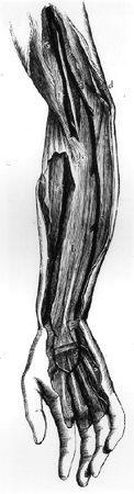
Image 252J , Image 497 , Image 498 Image 501
Anatomy Atlases. Image hosted by the site it comes from. Open the original page.
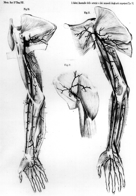
Image 252J , Image 497 , Image 498 Image 501
Anatomy Atlases. Image hosted by the site it comes from. Open the original page.
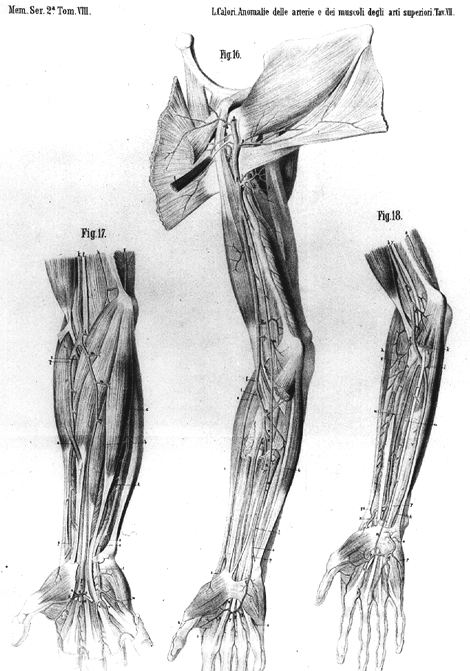
Image 252J , Image 497 , Image 498 Image 501
Anatomy Atlases. Image hosted by the site it comes from. Open the original page.
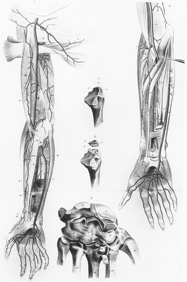
Image 252J , Image 497 , Image 498 Image 501
Anatomy Atlases. Image hosted by the site it comes from. Open the original page.
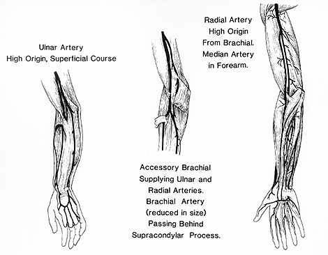
Image 252J , Image 497 , Image 498 Image 501
Anatomy Atlases. Image hosted by the site it comes from. Open the original page.
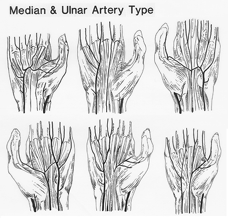
Image 252J , Image 497 , Image 498 Image 501
Anatomy Atlases. Image hosted by the site it comes from. Open the original page.
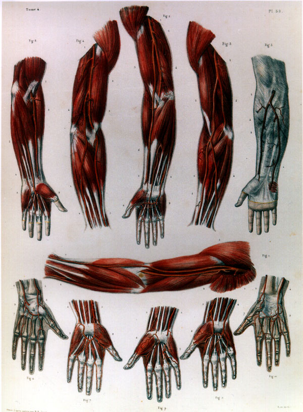
Image 252J , Image 497 , Image 498 Image 501
Anatomy Atlases. Image hosted by the site it comes from. Open the original page.
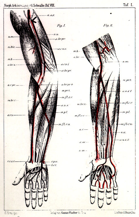
Image 252J , Image 497 , Image 498 Image 501
Anatomy Atlases. Image hosted by the site it comes from. Open the original page.
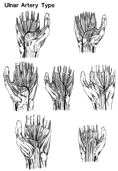
Image 252J , Image 497 , Image 498 Image 501
Anatomy Atlases. Image hosted by the site it comes from. Open the original page.
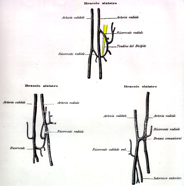
Image 252J , Image 497 , Image 498 Image 501
Anatomy Atlases. Image hosted by the site it comes from. Open the original page.
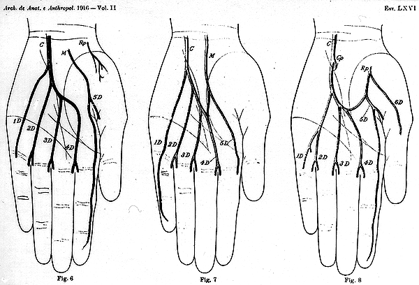
Image 252J , Image 497 , Image 498 Image 501
Anatomy Atlases. Image hosted by the site it comes from. Open the original page.
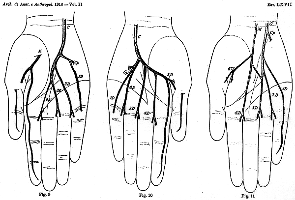
Image 252J , Image 497 , Image 498 Image 501
Anatomy Atlases. Image hosted by the site it comes from. Open the original page.
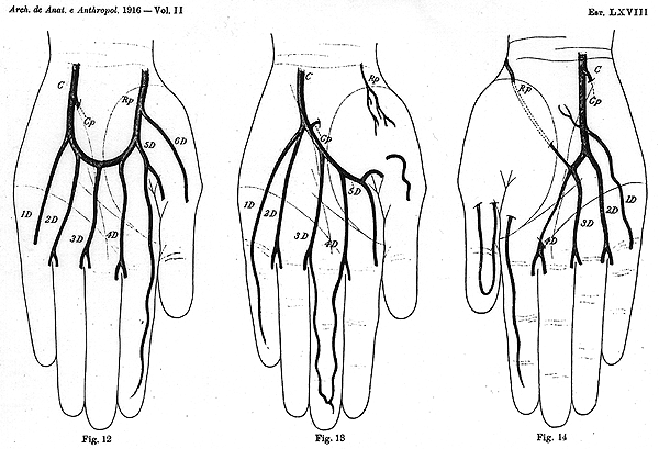
Image 252J , Image 497 , Image 498 Image 501
Anatomy Atlases. Image hosted by the site it comes from. Open the original page.
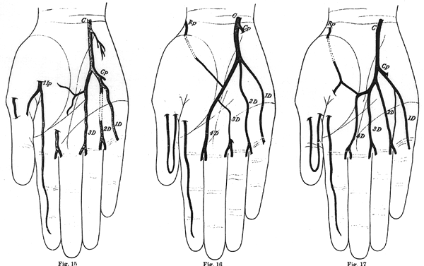
Image 252J , Image 497 , Image 498 Image 501
Anatomy Atlases. Image hosted by the site it comes from. Open the original page.
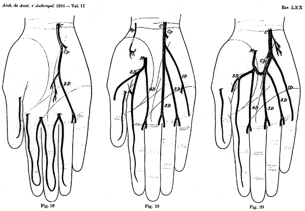
Image 252J , Image 497 , Image 498 Image 501
Anatomy Atlases. Image hosted by the site it comes from. Open the original page.
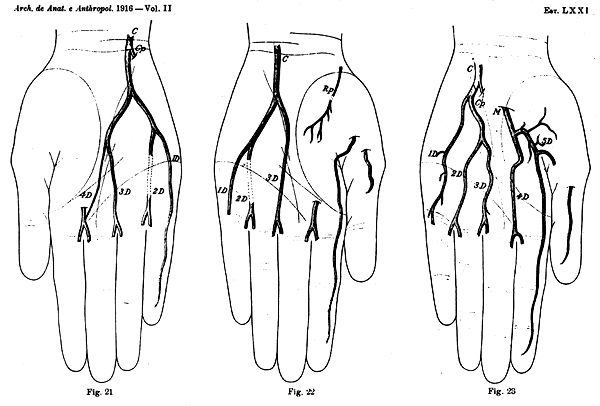
Image 252J , Image 497 , Image 498 Image 501
Anatomy Atlases. Image hosted by the site it comes from. Open the original page.
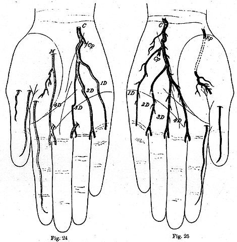
Image 252J , Image 497 , Image 498 Image 501
Anatomy Atlases. Image hosted by the site it comes from. Open the original page.
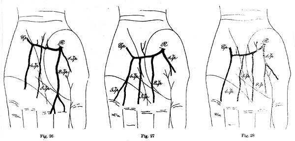
Image 252J , Image 497 , Image 498 Image 501
Anatomy Atlases. Image hosted by the site it comes from. Open the original page.
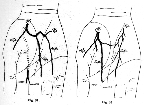
Image 252J , Image 497 , Image 498 Image 501
Anatomy Atlases. Image hosted by the site it comes from. Open the original page.
Deep
See Images 252A , 252B , 252C , 252D , 252E , 252F , 252G , 252H , 252I , 252J
Recurrent
Superficial
Anatomy Atlases. Image hosted by the site it comes from. Open the original page.
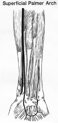
Posterior Interosseus
Anatomy Atlases. Image hosted by the site it comes from. Open the original page.
Posterior Interosseus
Anatomy Atlases. Image hosted by the site it comes from. Open the original page.
Posterior Interosseus
Anatomy Atlases. Image hosted by the site it comes from. Open the original page.
Posterior Interosseus
Anatomy Atlases. Image hosted by the site it comes from. Open the original page.
Posterior Interosseus
Anatomy Atlases. Image hosted by the site it comes from. Open the original page.
See Image 250
Median, superficial
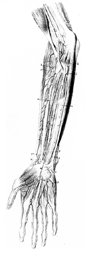
Anatomy Atlases. Image hosted by the site it comes from. Open the original page.
Riferimenti dell’opera originale
Elenco riportato come compare nella fonte, senza normalizzazione dei formati bibliografici.
- Anson, B.J., Ed. (1966) Morris' Human Anatomy, 12th ed., The Blakiston Division, McGraw-Hill Book Company, New York.
- Beaumanoir (de Brest), -. (1882) Présentations de pièces anatomiques: l'artère cubitale. Bull. de la Soc. Anatomique de Paris 57:319.
- Bergman, R.A., Thompson, S.A., Afifi, A.K. and F.A. Saadeh. (1988) Compendium of Human Anatomic Variation: Catalog, Atlas and World Literature. Urban & Schwarzenberg., Baltimore and Munich.
- Bianchi, L. (1943) Considerazioni sopra un caso di arteria brachio-ulnare superficiale. Anat. Anz. 94:73-88.
- Brême, G. (1899) Casuitischer Beitrage zur Kenntniss der Anomalien der Armarterien. Zeitschr. für Morphology und Anthropology 1:494.
- Chassaignac, -. (1834) Anomalie de l'artère cubitale. Bull. et Mém. de la Soc. Anatomique de Paris 1834:2.
- Cocchi, A. (1891) Contribuzione allo studio dell'anastomosi tra radiale e cubitale alla piegatura del cubito nella divisione prematura dell'arteria brachiale. Atti dell Accademia dei Fisiocritici in Siena. Ser. 4, 3:247-261.
- Dean, M.H. (1880) Arterial anomalies. The Medical Record, N.Y. 17:384.
- Demeaux, -. (1843) Anomalie des artères de l'avant-bras. Bull. et Mém. de la Soc. Anatomique de Paris 1843:72.
- Demeaux, -. (1843) (Superficial cubital and radial arteries) Bulletins et Mem. de la Société Anatomique de Paris XVIII(2):72.
- Gruber, W. (1867) über die Arteria mediana antibrachii superficialis, Arteria ulnaris antibrachii superficialis und Duplicität der arteria ulnaris. Arch. Anat. Physiol. Wissen. Med. 1867:668-687
- Gruber, W. (1871) Duplicität der Arteria ulnaris - neuer Fall. Arch. Anat. Physiol. Wissen. Med. 1871:286-296.
- Guerra, P. (1887) Anomalie rinvenute in cadaveri di delinquenti e di normali. Ateneo Medico Parmense 1:278-272.
- Guerrier, Y. and R. Paleirac. (1951) Le tronc artèriel antibrachial. Assoc. Anatomistes Comptes Rendus 38:566-575.
- Guyon, F. (1861) Note sur une disposition anatomique propre a la face antérieure de la région du poignet et non encore décrite. Bulletins et Mémoires. Société Anatorniques de Paris. 6:184-186.
- Hazlett, J.W. (1949) The superficial ulnar artey with reference to accidental intra-arterial injection. Canad. Med. Assoc. J. 61:289-293.
- Heuston, F.T. (1885) Notes on some anatomical anomalies. Trans. - Royal Academy of Medicine in Ireland 3:453-456.
- Maruyama, K. (1944) Seltene Varietät der Arterien der oberen Extremität bei einem Japaner (A. brachialis superficialis lateralis inferior, A. antebrachialis superficialis mediano-ulnaris, Arcus volaris superficialis vom Typus mediano-ulnaris). Folia Anat. Jpn. 22:551-567.
- Pabst, R. and H. Lippert. (1968) Beiderseitiges Vorkommen von A. brachialis superficialis, A. ulnaris superficialis, und A. mediana. Anat. Anz. 123:223-226.
- Poynter, C.W.M. (1922) Congenital anomalies of the arteries and veins of the human body with bibliography. The University Studies of the University of Nebraska 22:1-106.
- Radzikowski, A. and P. Szulczyk. (1972) The radial recurrent and ulnar recurrent arteries. Folia Morphol. 31:109-116.
- Ramsay, A. (1813) Account of unusual conformation of some muscles and vessels. Edinburgh Medical and Surgical Journal 8:281-283.
- Schwalbe, E. (1889) Beitrag zur Kenntniss der Arterienvarietäten des menschlichen Arms. Morphologichen Arbeiten 8:1-47. Schwalbe, E. (1898) Beitrag zur Kenntniss der Arterienvarietäten des menschlichen Arms. Morphologische Arbeiten 8:1-47.
- Souligoux, Ch. (1895) Anomalie de l'artère cubitale. Bull. et Mém. de la Soc. Anatomique de Paris 1895:657.
- Stocquart, A. (1887) Les anomalies de l'artère cubitale. Archives de Médecine et de Chirurgie Pratiques 2:85-86.
- Tedeschi, -., Torraca, -. and -. Pavone. (1907) Anomalia delle arterie dell'antibraccio e del nervo mediano. Giorn. Internaz. delle Scienze Mediche 29:315-317.
- Weathersby, H.T. (1956) Unusual variation of the ulnar artery. Anat. Rec. 124:245-248.
- Winslow, R. (1883) A study of the malformations, variations, and anomalies of the circulatory apparatus in man. Annals of Anatomy and Surgery 7:21-22.
Anatomical classification
- Systems:
- Cardiovascular system
Content provenance
Adaptation
Adaptation of «Ulnar Artery — Illustrated Encyclopedia of Human Anatomic Variation: Opus I: Muscular System» by Ronald A. Bergman, Adel K. Afifi, Ryosuke Miyauchi. Source: https://www.anatomyatlases.org/AnatomicVariants/Cardiovascular/Text/Arteries/Ulnar.shtml Original work distributed under the CC BY-NC-SA 4.0 licence (https://creativecommons.org/licenses/by-nc-sa/4.0/deed.it). Adaptation by Redazione AIVA. Changes from the original: Testo originale inglese riprodotto integralmente e riorganizzato nel formato scheda del portale (sintesi, testo, riferimenti), senza modificarne le affermazioni. La traduzione italiana non è ancora stata eseguita, e la pagina lo dichiara. Le figure non sono state copiate: sono collegate alla pagina d’origine. This publication is not approved or endorsed by the authors of the original work.
Licence
This content is distributed under the licence CC BY-NC-SA 4.0. Licences and attribution