Encyclopedia entry
Celiac Trunk Arteries
Abstract
The typical three-branched celiac trunk occurred in 86% of 756 cases studied. The celiac trunk may be absent (0.4%, Poynter); the branches usually arising from it then arise from the aorta. In one case, the splenic gave rise to the left gastric, and the common hepatic originated from the superior mesenteric. The trunk may be shorter or longer than usual. In the latter case, the branches commonly arise separately from the trunk instead of radiating from a single point.
The typical three-branched celiac trunk occurred in 86% of 756 cases studied.
The celiac trunk may be absent (0.4%, Poynter); the branches usually arising from it then arise from the aorta. In one case, the splenic gave rise to the left gastric, and the common hepatic originated from the superior mesenteric.
The trunk may be shorter or longer than usual. In the latter case, the branches commonly arise separately from the trunk instead of radiating from a single point.
The trunk may have only two branches, usually the splenic and the common hepatic, more rarely the left gastric and splenic, or it may have more than three branches. The additional branch may be one of the inferior phrenics, a trunk common for the inferior phrenics or for a inferior phrenic and left gastric, a gastroduodenal (or an accessory gastroduodenal), a second left gastric or an accessory splenic artery, a superior mesenteric (1.4%, Poynter), a middle or accessory middle colic, a supreme pancreatic, or a dorsal pancreatic.
One of the usual branches of the celiac artery may be absent, and may be replaced by a stem common to the inferior phrenics, by the right middle suprarenal and the right gastroepiploic, or more rarely by some other branch. One of the branches of the celiac trunk may give rise to the superior mesenteric artery.
The celiac, superior mesenteric, and inferior mesenteric may be joined together by one continuous longitudinal anastomosis.
Cases of "celiac trunk" in the form of a splenogastric trunk or a hepatosuperior mesenteric trunk have been reported.
A branch of the celiac trunk may pass through the esophageal hiatus of the diaphragm to supply the lower medial quarter of the right lung.
The following table illustrates the percentage frequency of variations commonly encountered (from Branco via Poynter):
Name
Specimens
Celiac Trunk complete
Celiac Trunk incomplete
Celiac-Mesenteric Trunk
No Trunk
Rossi
102
84%
11%
2.5%
2.5%
Leriche
55
84
9
1
0
Descomps
50
88
12
0
0
Branco
50
89
8
2
0
Picquand
50
82
14
2
2.0
Lipschutz
83
72
25
3
0
Eaton
206
90
9
1
0
Poynter
160
89
9
2
0
Totals
756
86%
11.8%
1.4%
0.4%
Dupuis and Barnay (1874) reported a celiac trunk giving rise to five arteries: 1) left gastro-epiploic, 2) splenic, 3) right gastro-epiploic, 4) hepatic and 5) inferior phrenic.
A celiac artery may give rise to an esophageal artery or a pulmonary artery.
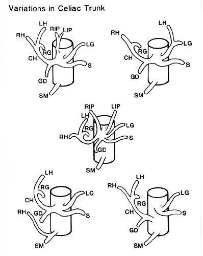
Image 333A
Anatomy Atlases. Image hosted by the site it comes from. Open the original page.
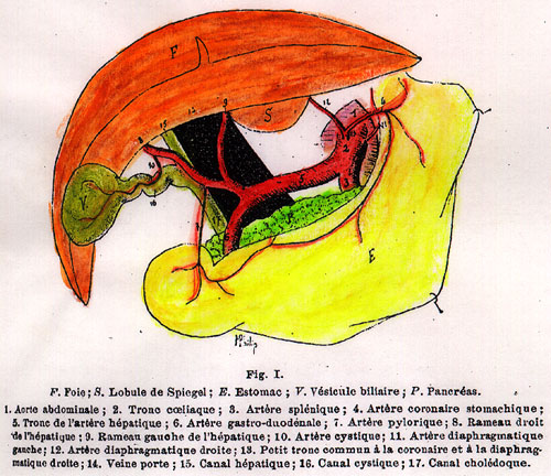
Image 333A
Anatomy Atlases. Image hosted by the site it comes from. Open the original page.
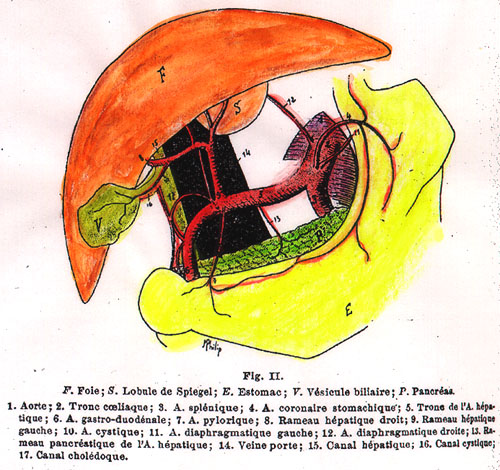
Image 333A
Anatomy Atlases. Image hosted by the site it comes from. Open the original page.
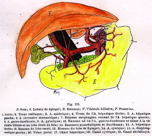
Image 333A
Anatomy Atlases. Image hosted by the site it comes from. Open the original page.
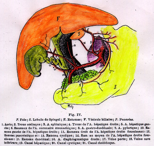
Image 333A
Anatomy Atlases. Image hosted by the site it comes from. Open the original page.
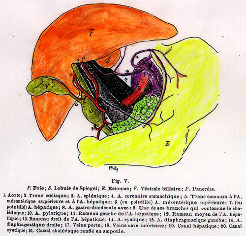
Image 333A
Anatomy Atlases. Image hosted by the site it comes from. Open the original page.
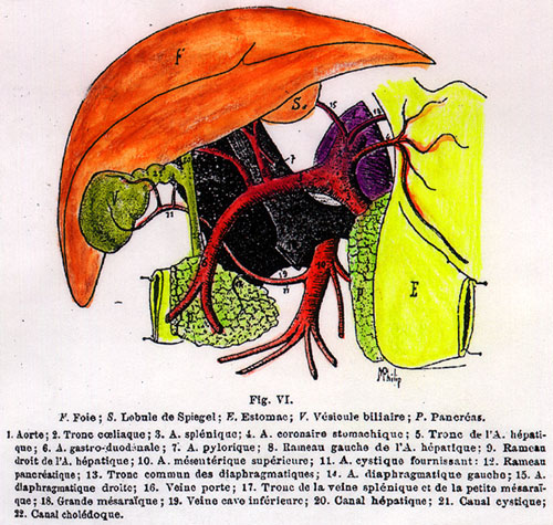
Image 333A
Anatomy Atlases. Image hosted by the site it comes from. Open the original page.
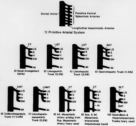
Image 333A
Anatomy Atlases. Image hosted by the site it comes from. Open the original page.
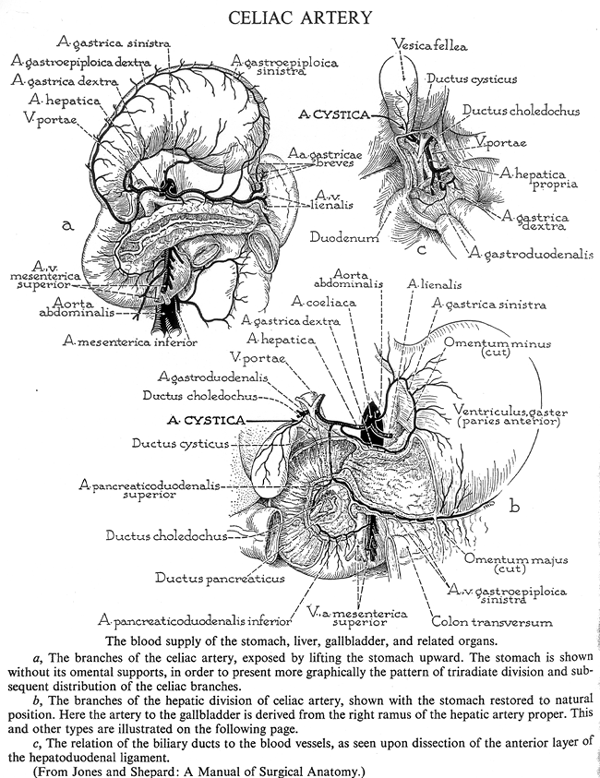
Image 333A
Anatomy Atlases. Image hosted by the site it comes from. Open the original page.
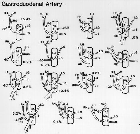
Gastroduodenal
Right Gastroepiploic
Anatomy Atlases. Image hosted by the site it comes from. Open the original page.
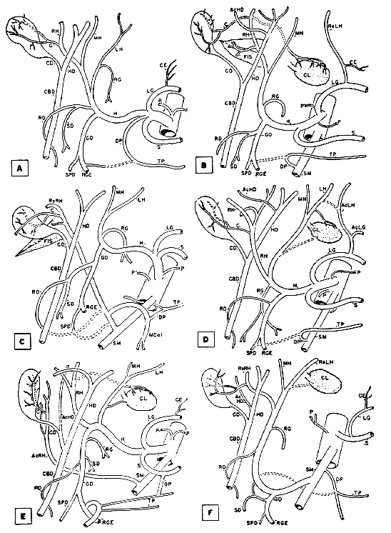
Right Gastroepiploic
Anatomy Atlases. Image hosted by the site it comes from. Open the original page.
Right Gastroepiploic
Anatomy Atlases. Image hosted by the site it comes from. Open the original page.
See ,
Anatomy Atlases. Image hosted by the site it comes from. Open the original page.
Dorsal Pancreatic
See Image 101
Splenogastric Trunk
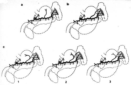
Hepatogastric Trunk
Anatomy Atlases. Image hosted by the site it comes from. Open the original page.
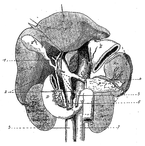
Hepatomesenteric Trunk
Anatomy Atlases. Image hosted by the site it comes from. Open the original page.
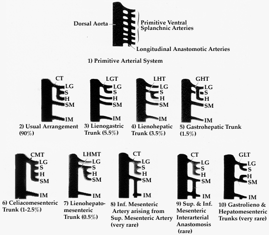
Lienogastric Trunk, Lienohepatic Trunk, and Lienohepatomesenteric Trunk
Anatomy Atlases. Image hosted by the site it comes from. Open the original page.
See Images 237, 333
Retroduodenal Course
See ,
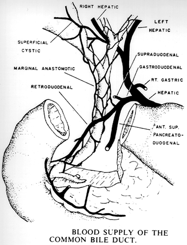
Anatomy Atlases. Image hosted by the site it comes from. Open the original page.
Gastrohepatic Trunk, Gastrolienal Trunk
See Images 237, 333
Gastrosplenic Trunk (of Splenic)
See ,
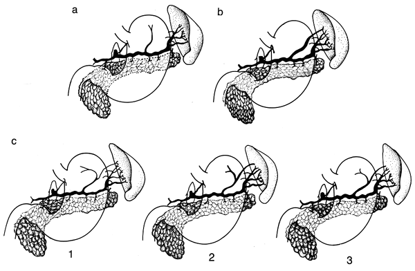
Anatomy Atlases. Image hosted by the site it comes from. Open the original page.
Riferimenti dell’opera originale
Elenco riportato come compare nella fonte, senza normalizzazione dei formati bibliografici.
- Augustyniak, E.A. (1965) A rare case of absence of the celiac artery. Folia Morphol., Warsaw 24:411-412.
- Bergman, R.A., Thompson, S.A., Afifi, A.K. and F.A. Saadeh. (1988) Compendium of Human Anatomic Variation: Catalog, Atlas and World Literature. Urban & Schwarzenberg, Baltimore and Munich.
- Bergmann, L. (1933) über einige seltene Anomalien der Arteria coeliaca. Z. Anat. Entwicklungsgesch. 101:525-533.
- Bogomolova, A.P. (1958) Arterial arches in the area of the pancreas head in man. Arch. Anat., Moskow 35:60-63.
- Caix, M. (1959) Variations topographiques des pédicules hépatiques et spléniques. Assoc. Anatomistes (Nancy) 47:231-242.
- Calas, F., Martin, R., Bouchet, Y. and D. Polliak. (1955) Les artères de la tête du pancreas. Assoc. Anatomistes, Comptes Rendus 42:362-367.
- Calori, L. (1880) Di una inversione splancnica generale nell'uomo accompagnata da alcuni notabili dell capo con esso lei convenienti e da estranee anomalie. Mem. R. Accad. Sci. Istituto di Bologna S. 4., 2:597-622.
- Chadzypanagiotis, D. and L. Amerski. (1978) A rare case of the celiac trunk anomaly. Folia Morphol., Warsaw 37:401-405.
- Chérigié, E., Mellière, D., Bennet, J., Doyon, D. and J.C. Chenard. (1967) Anatomie radiologique de la vascularisation du pancréas. J. de Radiol. d'Electrologie et de Méd. Nucl. 48:346-352.
- Chiene, J. (1869) Complete obliteration of celiac and mesenteric arteries; the viscera receiving their blood supply through extra-peritoneal system of vessels. J. Anat. Physiol. 3:65-72.
- Chomet, -. (1897) Anomalie du tronc coeliaque. Bull. et Mém. de la Soc. Anatomique de Paris 1897:954.
- van Dorshe, H.H. and A. Dorn. (1966) Varietäten im Bereiche der A. coeliaca und an den Nierengefässen. Anat. Anz. 118:297-304.
- Dunn. L.A., Washburn, J.W. and J.H. Targett. (1890) Abnormalities observed in the dissecting room of Guy's Hospital during sessions 1888-1890. S. 3., 32:299-315.
- Dupuis, -. et -. Barnay. (1874) Anomalie des branches du tronc coeliaque, et notamment artère hépatique double. Lyon Medical 15:499
- Duval, J.M., Prevot, J. et J.P. Loescher. (1959) Un cas de tronc coeliaco-mésentérique donnant deux artères hépatiques. Assoc. Anatomistes, Comptes Rendus 46:231-236.
- Eaton, P.B. (1917) The coelic axis. Anat. Rec. 13:369-374.
- Edwards, L.F. (1941) The retroduodenal artery. Anat. Rec. 81:351-355.
- Falconer, C.W.A. and E. Griffiths. (1949-50) The anatomy of blood-vessels in the region of the pancreas. Br. J. Surg. 37:334-344.
- Fontaine, C., Libersa, C., Chambon, J.P. and M. Ribet. (1980) La vascularisation artèrielle de la queue du pancréas et les nécroses caudales pancreatiques après splénectomie. J. Chirur. (Paris) 117:672-682.
- George, R. (1935) Topography of the unpaired visceral branches of the abdominal aorta. J. Anat. 69:196-205.
- Gérard, G. et D. Cordonnier. (1914) Cas type de triplicité de l'artère hépatique. Bibliographie Anatomique 24:211-217.
- Gisel, A. (1943) Eine seltene Variation im Verästelungstypus der arteria coeliaca. Ein Beitrag zur Frage der "ventralen Längsanastomose." Anat. Anz. 94:209-220.
- Grieg, H.W., Anson, B.J. and S.S. Coleman. (1951) The inferior phrenic artery. Types of origin in 850 body-halves and diaphragmatic relationship. Q. Bull. Northwestern University Medical School 25:345-350.
- Guibé, M. (1901) Sur la ligature de l'artere coronaire stomachique à propos d'une anomalie de cette artère. Bulls. et Méms. de la Soc Anatomique de Paris 3:212-214.
- Hecht, V. (1905) über einen Fall von Kollateralkreislauf im Gebiet der Arteria coeliaca. Anat. Anz. 26:570-576.
- Horrocks, P., White, W.H. and W.A. Lane. (1884) An account of the abnormalities observed in the dissecting room during the Winter session 1882-1883. Guy's Hospital reports S. 3., 27:39-48.
- Jacques, P. (1895) Quelques anomalies artérielles (sous clavière droite, tronc cliaque, poplitée). Bull. et Mem. de la Soc Anatomique de Paris 70:252-255
- Kosinski, C. (1928) Quelques observations sur les rameaux du tronc cliaque et des artères mésentèriques chez l'homme basée sur l'examen de 55 pièces anatomiques à Wilmo. Assoc. Anatomistes, Comptes Rendus 23:241-260.
- Kostinovitch, L.I. (1937) A case of simultaneous occurrence of a number of variations of the visceral branches of the abdominal aorta. Anat. Rec. 67:399-403.
- Lipshutz, B. (1917) A composite study of the coeliac axis artery. Ann. Surg. 65:159-169.
- Merklin, R.J. and N.A. Michels. (1958) The variant renal and suprarenal blood supply with data on the inferior phrenic, ureteral and gonadal arteries. A statistical analysis based on 158 dissections and review of the literature. J. Internat. College Surg. 29:41-76.
- Minne, J., Voisid, C. and C. Libersa. (1953) A propos de l'irragation artérielle de l'estomac et du bulbe duodenal. Assoc. Anatomistes, Comptes Rendus. 40:396-408.
- Nowak, M. (1968) Variation of the coeliac trunk in man. Folia Morphol., Warsaw 27:51-69.
- Olsen, L.L. and R.T. Woodburne. (1954) The vascular relations of the pancreas. Surg., Gynecol. Obstet. 99:713-719.
- Petrén, T. (1929) Die Arterien und Venen des Duodenums und des Pancreaskopfes beim Menschen. Z. Anat. Entwicklungsgesch. 90:234-277.
- Pierson, J.M. (1943) The arterial blood supply of the pancreas. Surg., Gynecol. Obstet. 77:426-432.
- Piquand, G. (1910) Recherches sur l'anatomie du tronc coeliaque et de ses branches. Bibliographie Anatomique 19(4): 159-201.
- Pitzorno, M. (1920-21) Morfologia delle arterie del pancreas. Arch. Ital. Anat. Embriol. 18:1-48.
- Poynter, C.W.M. (1922) Congenital anomalies of the arteries and veins of the human body with bibliography. The University Studies of the University of Nebraska. 22:1-106.
- Roberts, W.H. and P. Engen. (1978) Anomalous blood supply to the upper abdominal organs secondary to atresia of the celiac trunk. Anat. Anz. 143:396-398.
- Ross, J.A. (1950) The surgical significance of the intestinal vascular patterns. Edinburgh Med. J. 57:572-579.
- Sangari, S.K. and Mtui, E.P. (2017) Hepatosplenic and left hepatogastric trunks as variant arteries of foregut – A case report. International Journal of Anatomical Variations (2016) 9: 50–52
- Schubel, A.L. (1955-56) Abnormer Verlauf der 10. Interkostalarterie in Verbindung mit seltenen Gefässvarietäten im Bereich der A. coeliaca. Anat. Anz. 102:279-286.
- Spina, G.P., Galeotti, F., Opocher, E., Santambrogio, R. and G. Pezzuoli. (1983) Le variations de l'artère hépatique dans la chirurgie de l'hypertension portale. J. Chir. (Paris) 120 (No. 11):647-649.
- Swigart, LaV.L., Siekert, R.G., Hambley, W.C. and B.J. Anson. (1950) The esophageal arteries. An anatomic study of 150 specimens. Surg., Gynecol. Obstet. 90:234-243.
- Tallmadge, G.K. (1939) On the fixity of the gastroduodenal artery. Anat. Anz. 88:161-167.
- Tandler, J. (1904) über die variatäten der Arteria coeliaca und deren Entwicklung. Anat. Hefte. 25:473-499.
- Thane, -. (1888) Obliteration of the coeliac axis. Proc. Anat. Soc. Great Britain and Ireland. J. Anat. Physiol. 22:xvii-xviii.
- Wilkie. D.P.D. (!911) The blood supply of the duodenum, with special reference to the supraduodenal artery. Surg., Gynecol. and Obstet. 13:399-405.
- Wilmer, H.A. (1941) The blood supply of the first part of the duodenum. With description of the gastroduodenal plexus. Surgery 9:679-687.
- Woodburne, R.T. and L.L. Olsen. (1951) The arteries of the pancreas. Anat. Rec. 111:255-270.
- Zagorsky, P. (1830) Abnormitas quoad ortum et numerum quarundam arteriarum. Memoires de la Academie Imperiale des Sciences de St, Petersbourg. 1830, (Series 7): 201-204.
- Ziegler, H.R. (1942) Excision of the head of the pancreas for carcinoma with studies of its blood supply. Surg., Gynecol. Obstet. 74:137-145.
- Zientarski, B. and A. Kaszuba. (1975) A rare varieation of the celiac trunk in the form of a gastrolienal and hepatomesenteric trunk. Folia Morphol. Warsaw 34:403-406.
- Zwerina, H. and S. Poisel. (1966) über eine Anastomose zwischen dem Truncus coeliacus, der Arteria mesenterica inferior mit anderen Varietäten der unpaarigen und paarigen Äste der Bauchaorta bei einem Individuum. Anat. Anz. 119:427-435.
Anatomical classification
- Systems:
- Cardiovascular system
Content provenance
Adaptation
Adaptation of «Celiac Trunk Arteries — Illustrated Encyclopedia of Human Anatomic Variation: Opus I: Muscular System» by Ronald A. Bergman, Adel K. Afifi, Ryosuke Miyauchi. Source: https://www.anatomyatlases.org/AnatomicVariants/Cardiovascular/Text/Arteries/CeliacTrunk.shtml Original work distributed under the CC BY-NC-SA 4.0 licence (https://creativecommons.org/licenses/by-nc-sa/4.0/deed.it). Adaptation by Redazione AIVA. Changes from the original: Testo originale inglese riprodotto integralmente e riorganizzato nel formato scheda del portale (sintesi, testo, riferimenti), senza modificarne le affermazioni. La traduzione italiana non è ancora stata eseguita, e la pagina lo dichiara. Le figure non sono state copiate: sono collegate alla pagina d’origine. This publication is not approved or endorsed by the authors of the original work.
Licence
This content is distributed under the licence CC BY-NC-SA 4.0. Licences and attribution