Voce enciclopedica
Femoral Vein
Abstract
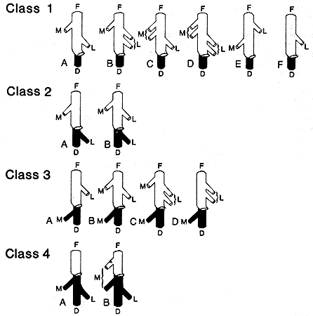
The femoral vein may be doubled in part of, or throughout its length. It may split into two and encircle the femoral artery. It occasionally passes through the adductor magnus above the femoral artery and runs independently from the artery until it joins the profunda vein to form the common femoral vein. The femoral may run with the sciatic nerve and pierce the the adductor magnus at the level of the lesser trochanter. When this occurs, the femoral artery is usually small, and the inferior gluteal (sciatic) artery is the primary (developmental) vessel of the thigh.
The femoral vein may be doubled in part of, or throughout its length.
It may split into two and encircle the femoral artery.
It occasionally passes through the adductor magnus above the femoral artery and runs independently from the artery until it joins the profunda vein to form the common femoral vein.
The femoral may run with the sciatic nerve and pierce the the adductor magnus at the level of the lesser trochanter. When this occurs, the femoral artery is usually small, and the inferior gluteal (sciatic) artery is the primary (developmental) vessel of the thigh.
A plexus forming a femoral vein has been reported.
The obturator may communicate with the femoral vein.

Lateral Femoral Circumflex and Medial Femoral Circumflex
Anatomy Atlases. Immagine ospitata dal sito d’origine. Apri la pagina originale.
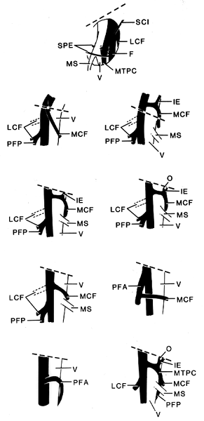
Lateral Femoral Circumflex and Medial Femoral Circumflex
Anatomy Atlases. Immagine ospitata dal sito d’origine. Apri la pagina originale.
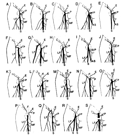
Lateral Femoral Circumflex and Medial Femoral Circumflex
Anatomy Atlases. Immagine ospitata dal sito d’origine. Apri la pagina originale.
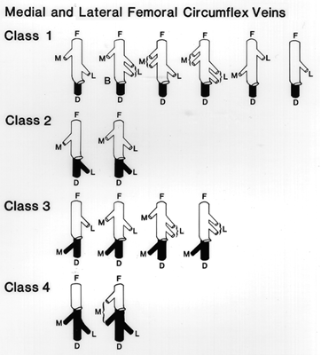
Superficial
Anatomy Atlases. Immagine ospitata dal sito d’origine. Apri la pagina originale.
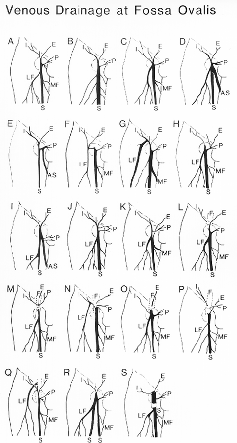
Superficial
Anatomy Atlases. Immagine ospitata dal sito d’origine. Apri la pagina originale.
Superficial
Anatomy Atlases. Immagine ospitata dal sito d’origine. Apri la pagina originale.
See Image 70
Riferimenti dell’opera originale
Elenco riportato come compare nella fonte, senza normalizzazione dei formati bibliografici.
- Arnold, G. von. (1968) Seltene Gefäss- und Muskelanomalien am Bein. Anat. Anz. 123:471-475.
- Bergman, R.A., Thompson, S.A., Afifi, A.K. and F.A. Saadeh. (1988) Compendium of Human Anatomic Variation: Catalog, Atlas, and World Literature. Urban & Schwarzenberg, Baltimore and Munich.
- Blair, Jr., C.B. and K. Nandy. (1965) Persistence of the axis artery of the lower limb. Anat. Rec. 152:161-172.
- Charles, C.M., Finley, T.L., Baird, R.D. and J.S. Cope. (1930) On the termination of the circumflex veins of the thigh. Anat. Rec. 46:125-132.
- Daseler, E.H., Anson, B.J., Reimann, A.F. and L.E. Beaton. (1946) The saphenous veinous tributaries and related structures in relation to the technique of high ligation. Surg., Gynecol. Obstet. 83:53-63.
- Edwards, E.A. and J.D. Roebuck. (1947) Applied anatomy of the femoral vein and its tributaries. Surg., Gynecol. Obstet. 85:547-557.
- Gruber, W. (1872) Anomalie Mündung der Vena saphena parva in die Vena cruralis. Arch. Pathol. Anat. Physiol. Klin. Med. 54:189.
- Gruber, W. (1867) Anomale Mündung der Ven saphena parva in die Vena cruralis. Archiv. f. Pathologische Anatomie u. Physiologie u. f. Klinische Medicin 54:189.
- Krahn, V. and H. Luzius. (1977) Besondere Lagebeziehungen kleiner Arterien zur Vena saphena magna im Bereich ihrer Einmündung in die Vena femoralis. Anat. Anz. 141:285-291.
- Le Dentu, -. (1867) Nouvelles recherchés sur la circulation veineuse de la jambe et du pied. Bulletins et Mem. de la Société Anatomique de Paris XLII(12):677-680.
- Nuzzi, O. (1943) Studio anatomica su particolari rapporti fra vene e arteria femorale nel settore medio della coscia. Archiv. Ital. Anat. Embriol. 48:1-21
- Poynter, C.W.M. (1922) Congenital Anomalies of the arteries and veins of the human body with bibliography. The University Studies of the University of Nebraska. 22:1-106.
Classificazione anatomica
- Sistemi:
- Sistema cardiovascolare
Provenienza del contenuto
Adattamento
Adattamento di «Femoral Vein — Illustrated Encyclopedia of Human Anatomic Variation: Opus I: Muscular System» di Ronald A. Bergman, Adel K. Afifi, Ryosuke Miyauchi. Fonte: https://www.anatomyatlases.org/AnatomicVariants/Cardiovascular/Text/Veins/Femoral.shtml Opera originale distribuita con licenza CC BY-NC-SA 4.0 (https://creativecommons.org/licenses/by-nc-sa/4.0/deed.it). Adattamento a cura di Redazione AIVA. Modifiche rispetto all’originale: Testo originale inglese riprodotto integralmente e riorganizzato nel formato scheda del portale (sintesi, testo, riferimenti), senza modificarne le affermazioni. La traduzione italiana non è ancora stata eseguita, e la pagina lo dichiara. Le figure non sono state copiate: sono collegate alla pagina d’origine. Questa pubblicazione non è approvata né avallata dagli autori dell’opera originale.
Licenza
Questo contenuto è distribuito con licenza CC BY-NC-SA 4.0. Licenze e attribuzioni