Voce enciclopedica
External Iliac Artery
Abstract
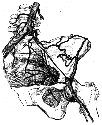
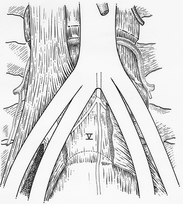
The external iliac may be longer or shorter than usual, resulting from the bifurcation of the common iliac above or below its usual point. When longer, it often takes a tortous course, making a partial loop or bend that may take it below the brim of the pelvis. The artery may be much smaller in diameter than usual; this may be the case when the femoral or main vessel of the lower limb arises from the inferior gluteal or other branch of the internal iliac. It then often terminates in profunda femoris.
The external iliac may be longer or shorter than usual, resulting from the bifurcation of the common iliac above or below its usual point. When longer, it often takes a tortous course, making a partial loop or bend that may take it below the brim of the pelvis.
The artery may be much smaller in diameter than usual; this may be the case when the femoral or main vessel of the lower limb arises from the inferior gluteal or other branch of the internal iliac. It then often terminates in profunda femoris.
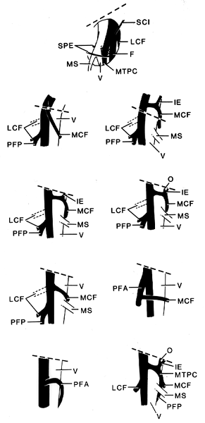
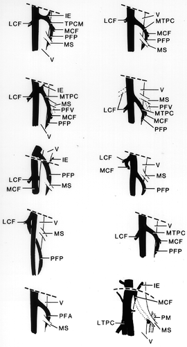
The external iliac may provide a large branch, such as the circumflex iliac or inferior epigastric, at a point higher than usual.
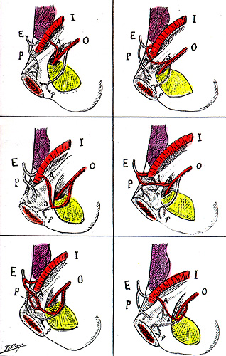
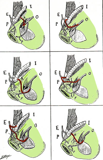
On occasion it gives rise to the obturator artey (1.1% of cases).
The artery may terminate as the deep femoral in cases in which the anastomoses between the inferior gluteal and popliteal are extensive.
In some cases, it gives rise to branches usually arising from the femoral, i.e., superficial external pudendal or profunda femoris.
In cases where the ischiadic artery persists as the main artery of the lower limb the external iliac is usually poorly developed.
Common Iliac
Anatomy Atlases. Immagine ospitata dal sito d’origine. Apri la pagina originale.
Common Iliac
Anatomy Atlases. Immagine ospitata dal sito d’origine. Apri la pagina originale.
Common Iliac
Anatomy Atlases. Immagine ospitata dal sito d’origine. Apri la pagina originale.
, , , , , , , See
Anatomy Atlases. Immagine ospitata dal sito d’origine. Apri la pagina originale.
Anatomy Atlases. Immagine ospitata dal sito d’origine. Apri la pagina originale.
Anatomy Atlases. Immagine ospitata dal sito d’origine. Apri la pagina originale.
Anatomy Atlases. Immagine ospitata dal sito d’origine. Apri la pagina originale.
Anatomy Atlases. Immagine ospitata dal sito d’origine. Apri la pagina originale.
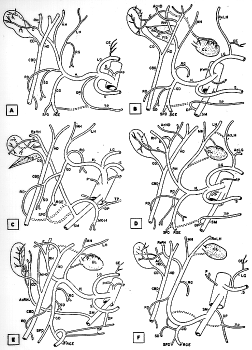
Anatomy Atlases. Immagine ospitata dal sito d’origine. Apri la pagina originale.
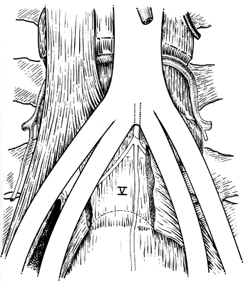
Anatomy Atlases. Immagine ospitata dal sito d’origine. Apri la pagina originale.
Riferimenti dell’opera originale
Elenco riportato come compare nella fonte, senza normalizzazione dei formati bibliografici.
- Anson, B.J., Ed. (1966) Morris' Human Anatomy, 12th ed. The Blakiston Division, McGraw-Hill Book Company, New York.
- Bergman, R.A., Thompson, S.A., Afifi, A.K. and F.A. Saadeh. (1988) Compendium of Human Anatomic Variation: Catalog, Atlas and World Literature. Urban & Schwarzenberg, Baltimore and Munich.
- Curtis, A.H., Anson, B.J., Ashley, F.L. and T. Jones. (1942) The blood vessels of the female pelvis in relation to gynecological surgery. Surg., Gynecol. Obstet. 75:421-423.
- Dschau, F. (1936-37) Eine bisher unbekannte Varietät der Arteria obturatoria und pudenda accessoria mit einer Varietät der A. circumflexa femoris medialis. Anat. Anz. 83:25-29.
- Lipshutz, B. (1918) A composite study of the hypogastric artery and its branches. Ann. Surg. 67:584-608.
- Pác, L., Hamplová, M. and O. Pelcová. (1977) An atypical case of arising of some parietal branches of the arteria iliaca imterna in man. Anat. Anz. 141:450-454.
- Poynter, C.W.M. (1922) Congenital anomalies of the arteries and veins of the human body with bibliography. University Studies of the University of Nebraska. 22:1-106.
Classificazione anatomica
- Sistemi:
- Sistema cardiovascolare
Provenienza del contenuto
Adattamento
Adattamento di «External Iliac Artery — Illustrated Encyclopedia of Human Anatomic Variation: Opus I: Muscular System» di Ronald A. Bergman, Adel K. Afifi, Ryosuke Miyauchi. Fonte: https://www.anatomyatlases.org/AnatomicVariants/Cardiovascular/Text/Arteries/IliacExternal.shtml Opera originale distribuita con licenza CC BY-NC-SA 4.0 (https://creativecommons.org/licenses/by-nc-sa/4.0/deed.it). Adattamento a cura di Redazione AIVA. Modifiche rispetto all’originale: Testo originale inglese riprodotto integralmente e riorganizzato nel formato scheda del portale (sintesi, testo, riferimenti), senza modificarne le affermazioni. La traduzione italiana non è ancora stata eseguita, e la pagina lo dichiara. Le figure non sono state copiate: sono collegate alla pagina d’origine. Questa pubblicazione non è approvata né avallata dagli autori dell’opera originale.
Licenza
Questo contenuto è distribuito con licenza CC BY-NC-SA 4.0. Licenze e attribuzioni