Voce enciclopedica
Deep Palmar Arch
Abstract
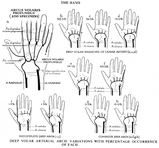
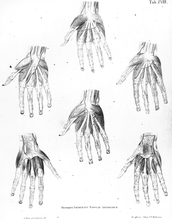
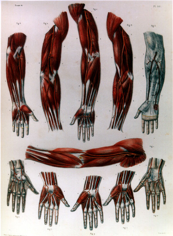
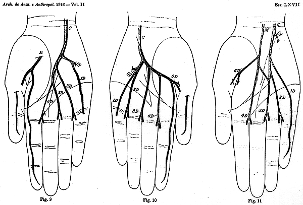
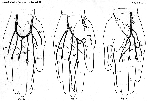
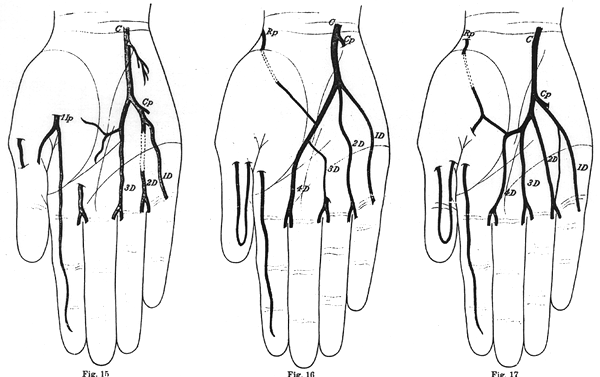
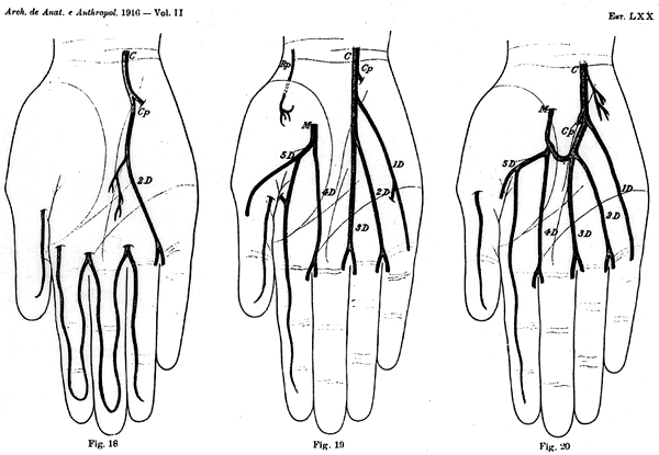
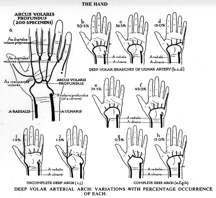
The deep palmar arch may be larger than usual, and its metacarpal branches may replace one or more of the common digital arteries by dividing at the cleft of the fingers into digital branches. The arch may be reinforced by enlarged posterior perforating branches from the radial and its branches on the back of the hand or by a large palmar interosseous. In some cases the radial joins the deep arch by passing through the second, instead of the first interosseous space.
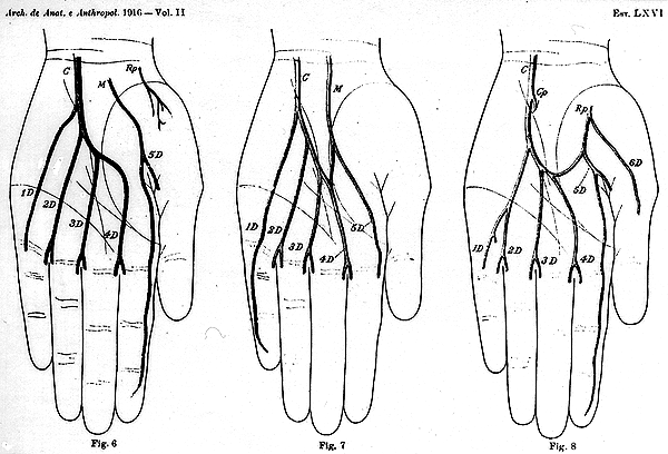
The deep palmar arch may be larger than usual, and its metacarpal branches may replace one or more of the common digital arteries by dividing at the cleft of the fingers into digital branches.
The arch may be reinforced by enlarged posterior perforating branches from the radial and its branches on the back of the hand or by a large palmar interosseous.
In some cases the radial joins the deep arch by passing through the second, instead of the first interosseous space.
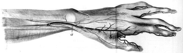
The princeps pollicis and radialis indicis may arise from the superficial palmar, or from a separate branch of the radial that passes through the first interosseous space.
When the radialis indicis originates from the superficial arch (13%) it is distributed to the radial side of the index finger.
The radialis indicis originates from the deep arch in 45% of cases, and the superficial arch in 13%; both arches contribute in 42%.
Of the 45% of individuals, radialis indicis and princeps pollicis arteries arise in common in 12% of cases. The princeps pollicis artery may arise from the radial artery in the palm, the superficial arch, or from a combination of both the deep and superficial arches.
The deep palmar arch is statistically less variable than the superficial palmar arch.
When absent (0.25%, Jaschtschinski), it is replaced by branches of the superficial palmar arch or by the dorsal system of arteries.
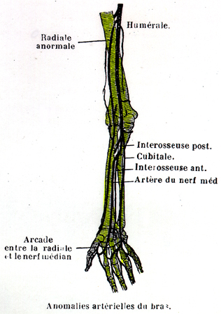
As can be readily ascertained, the arteries of the hand frequently vary from the expected or usual configuration. This occurs in two ways: 1) By far the greater number of deviations consist of a deficiency in either the radial or the ulnar system of arteries, accompanied by a corresponding increase in the other. The variation is much more commonly due to a deficiency in the superficial arteries with an increase supply from the deep set. 2) In the second and smaller group of variations a deficiency in one or another of the two systems is supplied, either by the enlargement of branches which descend in front of the limb, as the superficial volar (from the radial), or the median artery (from the anterior interosseous), or by the enlargement of an interosseous branch (from the radial) on the back of the hand.
Schaefer, et al., in Quain's Anatomy illustrated these general remarks as follows: " In the greater number of cases the superficial palmar arch is diminished, and gives off fewer digital branches than usual. Generally only one branch is wanting, viz., that which supplies the adjacent sides of the fore and middle fingers; but sometimes two or three branches are absent, or even all four, as when the ulnar artery, after giving branches to the short muscles of the little finger, ends in the deep palmar arch. In the last-mentioned case, which is rare, it is obvious that the superficial arch is altogether wanting."
"These various deficiencies in the superficial palmar arch and its branches are usually compensated by an enlargement of the deep arch, the palmar interosseous branches of which, being increased in size, divide at the clefts of the fingers, and form such collateral digital branches as are not derived from the usual source. But a defective superficial arch may, as before mentioned, be reinforced from other vessels, viz., from the superficial volar, from an enlarged median artery, or from a large dorsal interosseous branch."
"It sometimes, but less frequently, happens that the deep system of vessels is deficient, in which case the superficial arch may supply all the digital arteries to the thumb and fingers, one or more of these may be derived from the superficial volar, the median, or a dorsal interosseous artery."
"The superficial palmar arch has occasionally been seen doubled, the superficial part of the ulnar artery and the superficial volar branch of the radial being each divided into two communicating branches. Two of the digital arteries frequently arise by a common trunk." This ends the general summary from Quain's Anatomy, 1915.
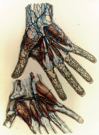
A common digital artery may penetrate a proper digital nerve creating a neural loop.
, , , , , , , , , , , , Image 252I , , , ,
Anatomy Atlases. Immagine ospitata dal sito d’origine. Apri la pagina originale.
Anatomy Atlases. Immagine ospitata dal sito d’origine. Apri la pagina originale.
Anatomy Atlases. Immagine ospitata dal sito d’origine. Apri la pagina originale.

Anatomy Atlases. Immagine ospitata dal sito d’origine. Apri la pagina originale.
Anatomy Atlases. Immagine ospitata dal sito d’origine. Apri la pagina originale.
Anatomy Atlases. Immagine ospitata dal sito d’origine. Apri la pagina originale.
Anatomy Atlases. Immagine ospitata dal sito d’origine. Apri la pagina originale.
Anatomy Atlases. Immagine ospitata dal sito d’origine. Apri la pagina originale.
Anatomy Atlases. Immagine ospitata dal sito d’origine. Apri la pagina originale.
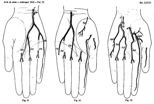
Anatomy Atlases. Immagine ospitata dal sito d’origine. Apri la pagina originale.
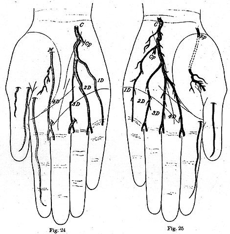
Anatomy Atlases. Immagine ospitata dal sito d’origine. Apri la pagina originale.
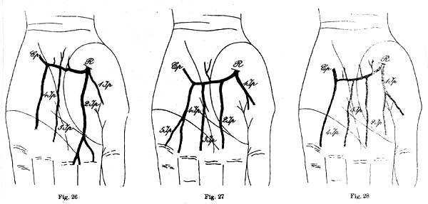
Anatomy Atlases. Immagine ospitata dal sito d’origine. Apri la pagina originale.
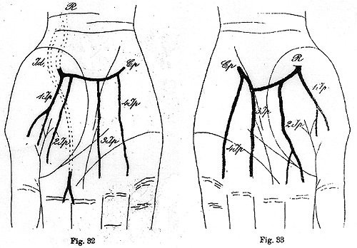
Anatomy Atlases. Immagine ospitata dal sito d’origine. Apri la pagina originale.

Anatomy Atlases. Immagine ospitata dal sito d’origine. Apri la pagina originale.
Anatomy Atlases. Immagine ospitata dal sito d’origine. Apri la pagina originale.
Anatomy Atlases. Immagine ospitata dal sito d’origine. Apri la pagina originale.
Princeps Pollicis and Radialis Indicis
Hand
Anatomy Atlases. Immagine ospitata dal sito d’origine. Apri la pagina originale.
, See Images 250 , 252A , 252B , 252C , 252D , 0252E , 252F , 252G , 252H , 252I , 252J
Anatomy Atlases. Immagine ospitata dal sito d’origine. Apri la pagina originale.
Riferimenti dell’opera originale
Elenco riportato come compare nella fonte, senza normalizzazione dei formati bibliografici.
- Barbosa Sueiro, M.B. (1916) Arcadas arteriais palmares. Arquivo de Anat. e Anthropol. 3:389-414.
- Barbosa Sueiro, M.B. (1917) Arcadas arteriais palmares. Arquivo de Anat. e Anthropol. 3:97-124.
- Bergman, R.A., Thompson, S.A., Afifi, A.K. and F.A. Saadeh. (1988) Compendium of Human Anatomic Variation: Catalog, Atlas and World Literature. Urban & Schwarzenberg, Baltimore and Munich.
- Coleman, S.S. and B.J. Anson. (1961) Arterial patterns in the hand based upon a study of 650 specimens. Surg., Gynecol. Obstet. 113:409-424.
- Henle, J. (1868) Handbuch der Anatomie des Menschen. Von Friedrich Vieweg und Sohn, Braunschweig.
- Jaschtschinski, S.N. (1897) Morphologie und Topographie des Arcus volaris sublimis und profundus des Menschen. Anat. Hefte 7:161-188.
- Latarjet, A. (1948) Testut's Traite d'Anatomie Humaine, 9th ed., G.Doin & Cie., Paris.
- Lippert, H. und Pabst, R. (1985) Arterial Variations in Man: Classification and Frequency. Bergmann Verlag, Munich.
- Mestdagh, H. (1980) Anatomie des Arcus volaris profundus beim Menschen. Anat. Anz. 147:180-187.
- Mozersky, D.J., Buckley, C.J., Hagood, Jr., C.O., Capps, Jr., W.F. and F.J. Dannemiller, Jr. (1973) Ultrasonic evaluation of the plantar circulation. A useful adjunct to radial artery cannulation. Am. J. Surg. 126:810-812.
- Poynter, C.W.M. (1922) Congenital anomalies of the arteries and veins of the human body with bibliography. The University Studies of the University of Nebraska. 22:1-106.
- Schaefer, E.A., Symington, J. and T.H. Bryce, Eds. (1915) Quain's Anatomy, 11th ed., Longmans, Green and Co., London.
- Rodet, -. (1875) Statistique sur l'artère radio-palmaire. Lyon Méd. 19:218-219.
- Schwalbe, E. (1898) Beitrag zur Kenntniss der Arterienvarietäten des menschlichen Arms. Morphologisches Arbeiten 8:1-47.
- Spinner, R.J., Varela, C.D. and J.R. Urbaniak. (1996) Digital nerve penetration by a digital artery in a patient with neurovascular symptoms: A case report. The Journal of Hand Surgery (Am) 21(6):1101-1103.
- Tandler, J. (1897) Zur Anatomie der Arterien der Hand. Anat. Hefte 7:263-282.
- Winslow, R. (1883) A study of the malformations, variations, and anomalies of the circulatory apparatus in man. Annals of Anatomy and Surgery 7:91-92.
Classificazione anatomica
- Sistemi:
- Sistema cardiovascolare
Provenienza del contenuto
Adattamento
Adattamento di «Deep Palmar Arch — Illustrated Encyclopedia of Human Anatomic Variation: Opus I: Muscular System» di Ronald A. Bergman, Adel K. Afifi, Ryosuke Miyauchi. Fonte: https://www.anatomyatlases.org/AnatomicVariants/Cardiovascular/Text/Arteries/ArchDeepPalmar.shtml Opera originale distribuita con licenza CC BY-NC-SA 4.0 (https://creativecommons.org/licenses/by-nc-sa/4.0/deed.it). Adattamento a cura di Redazione AIVA. Modifiche rispetto all’originale: Testo originale inglese riprodotto integralmente e riorganizzato nel formato scheda del portale (sintesi, testo, riferimenti), senza modificarne le affermazioni. La traduzione italiana non è ancora stata eseguita, e la pagina lo dichiara. Le figure non sono state copiate: sono collegate alla pagina d’origine. Questa pubblicazione non è approvata né avallata dagli autori dell’opera originale.
Licenza
Questo contenuto è distribuito con licenza CC BY-NC-SA 4.0. Licenze e attribuzioni